Icefish: one of the extremes in the animal kingdom aids ophthalmic research

25 March 2019 I Read more news
By Henrik Lauridsen, PhD, Department of Clinical Medicine, Aarhus University
Sight is one of our most important senses, and the retina of the eye is one of the most fascinating structures in humans and the animal kingdom generally.
The retina is one of the most energydemanding structures in the body, and a healthy retina requires a reliable and stable blood supply to meet its need for oxygen and nutrients.
A healthy retina has both external vessels in an anatomical structure – called choriocapillaris – on the outside of the retina, and retinal vessels on the inside. The fine network of blood vessels is perfectly adapted to match the oxygen supply required by the retina.
Disruption of this balance can cause dire complications, including severely impaired vision and even blindness.

In patients afflicted by wet age-related macular degeneration, weakening of the retina's outer layer causes the formation of new vessels – neovascularisation – behind the retina, and ingrowth or abnormal linkage of newly formed vessels in the outer layers of the retina. This results in an increase in fluid in the retina and impaired vision.
In patients with diabetic retinopathy, neovascularisation on the interior of the retina poses a risk of vision impairment, as the newly formed vessels tend to be fragile, and can easily rupture, which results in bleeding on the inside of the retina.
Given these risks in humans, it is astonishing that animals with extensive vascular ingrowth in the retina, do not, apparently, have the same problems.
Retinal structure is largely preserved in all animal species with a backbone (vertebrates), and by studying animals that deal with this problem perfectly, we can discover how neovascularisation of the retina can occur without any harmful effect. This situation is known
as the August Krogh principle, which states that for every medical problem ”...there will be some animal of choice or a few such animals on which it can be most conveniently studied”.
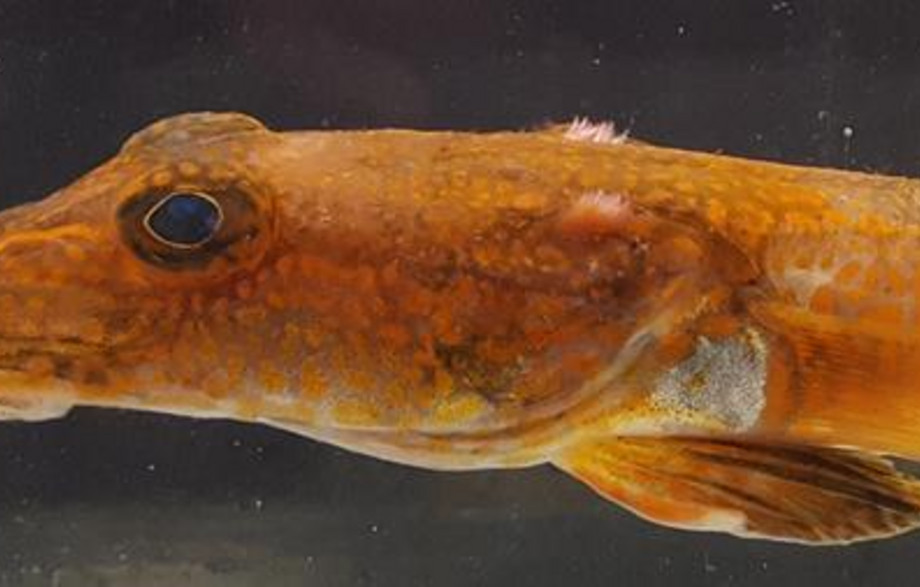
It turns out that the vertebrates with the largest ingrowth of retinal vessels are a family of Antarctic fishes named crocodile icefish or white-blooded fish. The name stems from the fact that these fish inhabit the icy waters of Antarctica's Southern Ocean, and are the only vertebrates that have none of the red oxygen-transporting pigment – haemoglobin – in their blood, which is why they are pale and have milky blood.

Without haemoglobin, their blood only has the capacity to transport 10% of the normal oxygen content, and to compensate for that, icefish have special adaptations in all oxygen- demanding organs and structures to ensure sufficient oxygen delivery. This is why icefish display extreme retinal neovascularisation during their development, with only a small percentage of the retinal surface not covered by ingrown blood vessels. But how can icefish withstand such extensive neovascularisation when we, and the animal models we traditionally study, cannot?
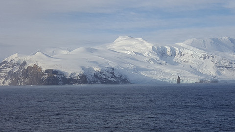
Research project for new insights"With funding from VELUX FONDEN, I am now able to use state-of-the-art scanning techniques to investigate this question on the the Antarctic Peninsula. The hope is that this research will provide a far deeper understanding of the adaptations that protect the fragile retina during the ingrowth of new blood vessels," says Henrik Lauridsen.
Henrik Lauridsen, Department of Clinical medicine, Aarhus University